©2017 World Federation of Pediatric Imaging - All rights reserved.


Disclaimer: the content offered on this site is prepared by individual authors or institutions; WFPI has no responsibility for it and does not endorse any products advertised therein. Material is for use only by trained medical personal licensed in their area of expertise in the location in which they practice. WFPI is an independent organization operating under the auspices of the Society for Pediatric Radiology (SPR)

SPR is a non-profit 501 (c) (3) organization in the United States.


Dr. Jennifer Nicholas
Chair, Digital Education and Social Media Committee

Webmaster


WFPI Mission
Communication and collaboration between pediatric imaging practitioners, via their organizations
Advocating for appropriate practices and resource allocation for children
Education
Patient safety, in particular radiation safety and protection
Outreach and training in lower resource settings
Research
Provision of information & resources


Publications

US research in lower resource settings

is featured in WFPI's mini-symposium "Outreach in the Developing World" in the Pediatric Radiology journal, issued May 2014

Click here


Relevant US publications

Other project partners

The American College of Radiology has funded project travel USA - South Africa.


ACRF International Outreach newsletter May 2014 


Preparatory steps taken

Protocols were tested in early December 2013 (Brian Garra, DB and Dr. Kara-Lee Pool, a UCLA resident). Dr. Pool travelled to South Africa in January 2014 to train a non-radiologist in the sweeps.

The gold standard radiologist was identified: Dr. Sabine Bélard, a PhD student in Cape Town with an accepted protocol and ethics approval for pediatric abdominal bedside ultrasound on HIV.

The research protocols and ethics amendments were coauthored by Drs. Kara-Lee Pool, Sabine Belard, Savvas Andronikou and Heather Zar, who represent the authors of the proposed research.

The Red Cross Hospital purchased a high-resolution, small footprint, sector transducer of 7.5 MHz for the project.


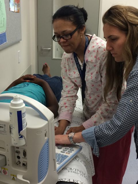
Dr. Kara-Lee Pool (UCLA) offering volume sweep training to Ms. Washiefa Isaacs, a nurse at the Red Cross Memorial Children's Hospital, Cape Town, South Africa, in preparation for the TB research project, January 2014. Dr. Pool is also documenting how this research project is presented to parents/primary carers so as to obtain consent for their children's enrolment. 


Further research

Drakenstein Trial, South Africa

In step with the research at Cape Town's Red Cross Hospital and WFPI's PoC ultrasound work in Malawi during an assessment/training trip in July 2014, Drs. Tracy Kilborn and Savvas Andronikou are also involved in a Red Cross/UCT project in the PAARL hospital, Drakenstein, run by Prof. Heather Zar (head of pediatrics, Red Cross War Memorial Children's Hospital, Cape Town). Clinicians are trained to diagnose pulmonary disease - pneumonia - at the point of care. UPDATE COMING SOON.


Dr. Tracy Kilborn training on ultrasound, Malawi, July 2014


.

Spreading the word....

WFPI outreach leader Prof. Savvas Andronikou speaking at the International Congress of Radiology, Dubai, September 2014


Volume sweep imaging and ultrasound

Project led for the WFPI by Savvas Andronikou, Dorothy Bulas & Ines Boechat


Project origins


WFPI is looking to become a pediatric partner for Imaging the World (ITW) which involves non-radiologists acquiring ultrasound images using anatomical landmarks on the skin and storing US sweeps as cine-loops in a standard format, to be read by radiologists at a distance (tele-reading). ITW has no pediatric protocols, so Drs. Dorothy Bulas & Brian Garra  (ITW technical co-founder) have prepared protocols for specific “management changing” conditions in resource-limited settings. They will include hydronephrosis, hydrocephalus and mediastinal lymphadenopathy (as a proxy for TB) volume sweep US protocols.


The end goal: use in rural clinics at the point of care (POC) for management changing decisions.

Roll out in South Africa

The TB volume sweep research protocols have received  approval and ethical clearance under the stewardship of Prof. Health Zar, Chief Paediatrician at Red Cross War Memorial Children's Hospital and Chair Professor of Pediatrics at the University of Cape Town. The project will roll out in the Red Cross War Memorial Children’s Hospital (image right), nested within a larger HIV-TB project.

A pilot study previously conducted in Johannesburg’s Chris Hani Baragwanath Hospital by Dr. Tsepo Moseme and Savvas Andronikou in 2013 has already shown that sonography of the mediastinum is a feasible, reliable, non-invasive method which is able to detect mediastinal lymphadenopathy in children with suspected primary pulmonary tuberculsos. As such it provides an alternative POC diagnostic test in the diagnosis of pediatric TB. This preliminary paper is accepted for publication in the Pediatric Radiology journal.

This WFPI TB volume sweep research project will determine quality of transferred information; compare volume sweep diagnosis against a gold standard US examination and compare the US detection of lymphadenopathy against plain radiography while taking other definitive diagnostic tests into account. There is also an associated TB MRI project run by Dr. Tanyia Pillay (SASPI) with Savvas Anrdonikou, which can serve as a gold standard for a proportion of the patients.

Patient enrolment:
Any child suspected of TB will receive all the standard diagnostic tests, followed by abdominal US performed by a trained sonographer. The patient will then receive  a mediastinal volume sweep, ITW-style, performed by a blinded non-radiologist healthcare  worker with no prior US experience, followed by a chest CXR, and in a subgroup, MRI of the chest.

Study objectives:

  • Demonstrate that a protocol incorporating volume sweep imaging technique of the mediastinum can be taught to an ultrasound naive volunteer with minimal training.
  • Evaluate the quality/interpretability of mediastinal sweeps performed by a minimal trained sweeper for the evaluation of mediastinal anatomy
  • Evaluate the quality/interpretability of mediastinal sweeps performed by a minimally trained sweeper for the evaluation of presence or absence of mediastinal lymphadenopathy in children with suspected TB. 
  • Compare the performance of the mediastinal sweeps done by a minimal-trained sweeper against the free hand bedside ultrasound performed by an ultrasound trained physician.
  • Compare the mediastinal ultrasound against the chest radiograph for presence of lymphadenopathy.

Renal and head US projects can also be performed in South Africa or alternatively in the USA, Europe or elsewhere as they do not require TB patients.

Progress updates

Report May 2015: 

Ms. Washiefa Isaacs, a Red Cross Hospital nurse with no previous ultrasound experience was trained for 8 hours to perform four mediastinal volume sweep imaging techniques on pediatric patients using a portable Mindray DP10 machine with a 5-8.5 MHz micro-convex array transducer. The four sweeps included: transverse and oblique sweeps of the suprasternal region, and transverse and longitudinal sweeps of the left lateral sternum.
Patients with suspected tuberculosis underwent diagnostic TB workup with the best available diagnostic tools and given the standard of care for TB treatment if indicated. Each patient received both VSI and free hand mediastinal ultrasound performed by an ultrasound trained pediatrician.  Patients were randomized to receive either VSI first or freehand mediastinal ultrasound first. While the ultrasound competent pediatrician was allowed to look at the screen while obtaining the images, Ms Isaacs was not trained in interpreting screen images, and therefore was instructed only to use anatomic landmarks to obtain her mediastinal VSI scans.
An independent technical reader reviewed the ability of the ultrasound naïve nurse to obtain the correct technical components of the sweeps.  Two pediatric radiologists and one ultrasound radiologist reviewed the sweeps.  Studies were reviewed for ability to identify normal anatomy as well as lymphadenopathy.

TECHNIQUE


There have been 37 pilot patients completed to date (June 2015).  Each patient was scanned using the four mediastinal VSI techniques by Ms Isaacs. The same patients also obtained the mediastinal free hand ultrasound performed by an ultrasound trained physician.  The results of the technical component of the study and the diagnostic component of the study are currently being analyzed.

Comparative statistical analysis will be used to compare VSI vs free hand ultrasound performed by the trained clinician: Chi Squared or Man Whitney U test.

Caution: Google (machine translation) is an imperfect art.
It is provided here as a service to our many colleagues worldwide.